How Nuclear Medicine Contributes to Diagnosing Lung Function Disorders
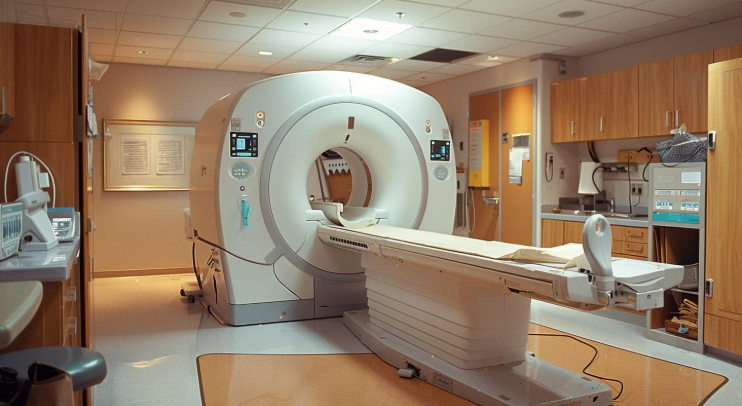
Nuclear medicine uses small amounts of radioactive materials to examine organ function and structure. Unlike other imaging techniques that show anatomy, nuclear medicine provides unique insights into physiological functions.. The process involves introducing a radiopharmaceutical into the body, which then accumulates in the specific organ or tissue being studied. Here is more information about this method and how it can help:
Delivering Radiotracers
Radiopharmaceuticals, also known as radiotracers, are central to nuclear medicine imaging of the lungs. Specialists prepare these compounds in a controlled setting, and they combine a radioactive isotope with a pharmaceutical that targets a specific biological process. They select the delivery method depending on the particular function being assessed.
Assessing Lung Ventilation
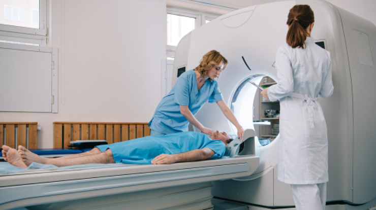
Nuclear medicine lung testing can evaluate the movement of air into and out of small air sacs. After you inhale a radioactive gas or aerosol, a gamma camera captures a series of images as the substance distributes throughout your airways. The resulting images show areas of the lungs that are receiving adequate air and identify regions with poor or obstructed airflow.
The procedure typically takes place over several minutes while you hold your breath at certain points; this allows for a detailed assessment of regional lung function. This detailed imaging technique is used to diagnose or monitor conditions such as pulmonary embolism or chronic obstructive pulmonary disease (COPD). It is a non-invasive and generally safe procedure, with minimal discomfort for the patient.
Checking Lung Perfusion
A lung perfusion scan assesses blood flow to the lungs. For this test, a radiotracer is typically administered through an intravenous injection, and it helps to trace the distribution of the substance within the body. The particles of the radiotracer are just large enough to get temporarily trapped in the small blood vessels of the lungs, allowing the gamma camera to visualize blood distribution. These nuclear medicine results reveal:
- Reveals how well blood circulates.
- Provides a map of pulmonary blood flow.
Medical providers typically complete this part of the examination quickly after the injection.
Diagnosing Conditions
Physicians use ventilation-perfusion (VQ) scans to diagnose specific lung conditions by comparing the results from both tests. A typical application is for detecting a pulmonary embolism (PE), which is a blood clot in the lung’s arteries. A mismatch between ventilation and perfusion is a classic sign of a PE. The images help pinpoint the location and size of any potential mismatches.
Radiologists can detect other conditions through VQ scanning. Chronic obstructive pulmonary disease (COPD) may present with matched defects, meaning both ventilation and perfusion are reduced in the same areas. These patterns assist your doctor in differentiating lung disorders, but careful analysis of combined images is necessary.
Quantitative data that radiologists derive from the scan provides objective metrics for lung function. The computer can calculate the ratio of ventilation to perfusion in different lung regions, and this data helps your physician. This information contributes to a comprehensive evaluation, especially when other imaging tests like a CT scan are not suitable or provide inconclusive results.
See also: HealthSciencesForumCom Team: Transforming Health Information for Everyone
Schedule Nuclear Medicine Sessions
Nuclear medicine procedures provide your healthcare provider with detailed insights into your lung function. These tests show how well your lungs are managing airflow and blood circulation, which helps in forming an accurate diagnosis. The specific findings from a VQ scan guide the next steps in your care plan. If you have been referred for this type of imaging, please contact our scheduling department to arrange your appointment.